|
Julian Moosmann Named Franklin Award Recipient for Development of Techniques for In vivo Analysis of Embryonic Development
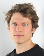
May 12, 2014 — The APS Users Organization has named Julian Moosmann as the winner of the 2014 Rosalind Franklin Young Investigator Award. The award recognizes Moosmann's work to develop methods that have made it possible to obtain time-lapse 3D images of cells and tissues in living vertebrate embryos. The methods will also apply more generally to imaging of materials that have low absorption contrast in conventional x-ray imaging. Moosmann, a physics Ph.D. student at Karlsruhe Institute of Technology (KIT), Karlsruhe, Germany, is cited as an "original, visionary thinker" who is equally willing to undertake cumbersome experimental work and elaborate numerical simulations. Nominators also commended his scientific maturity, his ability to master techniques from several fields and to work as a team player, and his deep understanding of the concepts and physics behind the experiments. Moosmann collaborated with his advisor, physicist Ralf Hofmann, and an international team of physicists, beamline scientists, and biologists from KIT, Northwestern University, and the APS to apply the new methods to image the movement of cells within an embryo of the African claw-toed frog (Xenopus laevis) over a period of about two hours. The results were published in Nature and the methods in Nature Protocols. The initial work was done at APS beamline 2-BM-B, and additional experiments at APS beamline 32-ID confirmed the results. The work recognized by this award provides an important new modality that could help answer fundamental questions in developmental biology. Although the much-studied Xenopus laevis has been the source of many insights about embryonic development, certain questions simply cannot be answered with current technologies. Light microscopes can't show cells inside the optically opaque embryos, and conventional absorption-based x-ray imaging is not sensitive enough, so phase-contrast techniques are needed. But it is still extremely challenging to capture cell movement within the developing embryo without killing it. Moosmann and Hofmann did not set out to revolutionize developmental biology. They work at ANKA, the synchrotron light source at KIT, and were interested in exploring the fundamental physics and mathematics of X-ray phase-contrast imaging, which exploits differences in a sample's refractive index—and the resulting phase changes in the transmitted x-rays—to create a signal. One of Moosmann's key advances was to modify the application of the contrast transfer function (CTF), a standard technique used in phase-contrast imaging. He wanted to improve the function's resolution at a greater distance from the sample, that is, at large z. "We decided to focus on propagation-based phase-contrast imaging because of the simplicity of the technique. The setup is free of additional optics and you need only one measurement; there's no scanning or stepping," Moosmann said.They also aimed for a method that would work at larger distances from the sample because in phase-contrast imaging the signal-to-noise ratio improves with distance. This point turned out to be an important advantage because it yields better resolution for less x-ray exposure, so embryos can live longer. It also permits the use of extended sample environments. The advance depended on close observation of the contrast transfer function. "There is a sinusoidal modulation in the Fourier transform of the intensity," Moosmann explained, "but when the variations in the phase increase, like what would happen as a sample becomes more complex, the contribution from the region around the maxima grew faster than that around the minima." This feature told him that information about the structure of the sample would be concentrated around the maxima. Also, the minima were always in the same place—up to point, at least. When he looked for where the function failed, Moosmann observed a critical transition phenomenon in which the minima shifted rapidly away from their previous positions. In a key step, he realized that below the critical transition, he could safely "crop out" small regions around the minima, leaving most of the information intact. Such critical transitions are familiar in condensed matter physics, so Moosmann and Hofmann borrowed a theoretical formalism from that field: they proposed that "phase" could be reinterpreted as a "quasiparticle." The same type of formalism is used as a shorthand to represent the vibration of atoms in a solid, for example. In technical terms, Moosmann explains, "There's a dispersion relation between image intensity and phase just as there is between energy and momentum in a solid state material." It was time for a real-world test. "I had a new algorithm and wanted to test it, so we got some fixed Xenopus embryos from Jubin Kashef, a biologist at KIT," Moosmann said. A proof-of-concept experiment at beamline ID19 at the European Synchrotron in Grenoble, France, was a success, yielding a resolution of about a micrometer. "Kashef was impressed with the detail that could be seen deep within the intact embryo," compared to what could be seen with light microscopy, said Moosmann, so they decided to undertake the ambitious step of an in vivo experiment using phase-contrast microtomography. It was a true trial-and-error scenario that demanded on-the-fly understanding of the physics and close collaboration with the beamline staff. "There are so many parameters; we just couldn't simulate them all," Moosmann said. Among them were the heat load, beam energy and bandwidth, exposure dose, pixel size, motion blur, scintillator thickness, number of tomographic projections, scheduling of embryonic development, time lapse between tomograms, and so on. It was a nerve-wracking juggling act. "On the very last scan of our beam time, the embryo survived 2 hours," Moosmann said—yielding enough data for the Nature paper. Optimizing the data collection was only the beginning. The image processing and data analysis were another hurdle. "I still had to work really hard to get a good-quality reconstruction of the data, especially since we were at the limits of that setup," Moosmann said. "The main work I'm doing is programming," he added. "There is so much data!" The result is a set of stunning time-lapse videos of a frog embryo in the gastrulation stage, during which the embryo forms three layers of cells that later become tissues and organs. Among the features shown in unprecedented detail is the formation of the archenteron, or primitive digestive tube. Other images include representations of velocity and direction of the movement of individual cells. The team achieved a resolution of about a micrometer, or about 1,000 times better than typical medical tomography. However, resolution of the technique is still limited by the coherence of the beam. With the coherence of the APS Upgrade, resolution and efficiency will substantially improve. By using a cone beam and local tomography, it should be possible to reach nanometer resolution, according to Moosmann. It has been quite a journey for the young German. "I didn't know much about biology. I had no clue when I started this what a blastopore was!" said Moosmann, who expects to complete his Ph.D. this summer. When he isn't learning an unfamiliar discipline or programming numerical simulations or writing algorithms, Moosmann loves climbing, playing squash, and enjoying really good food with his girlfriend, a chemist turned professional chef. "I'm a foodie," he admits. But it's clear that his passion of passions is thinking deeply about theory; his first degree (the German diploma) was in theoretical physics, also with Ralf Hofmann. "I've always had a big interest in theoretical concepts. After my diploma I wasn't yet a theoretician; that takes a long time. Now I'm somewhere in between a theoretician and an experimentalist. I'm interested in applying the theory to what you find at the beamline." |
| Further Reading |
|
Highlight
On in vivo imaging
On phase retrieval methods
|

